Research 2025
This study sought to evaluate and assess if any patient benefits were derived from the application of a specific physical therapy approach to the treatment of trigger points (TP’s).
Evaluation was performed using myotonometry (MyotonPRO Digital Palpation Device).
Physical therapy treatment used was MSTR® (McLoughlin Scar Tissue Release).
Measurement of Trigger Points Using Myotonometry
Pre and Post MSTR® treatment evaluation


Research 2023
This study sought to evaluate and assess if any patient benefits were derived from the application of MSTR® for the treatment of Cesarean Section scars.
Evaluation was performed using myotonometry (MyotonPRO Digital Palpation Device).
Measurement of C-section scars Using Myotonometry
Pre and Post MSTR® treatment evaluation
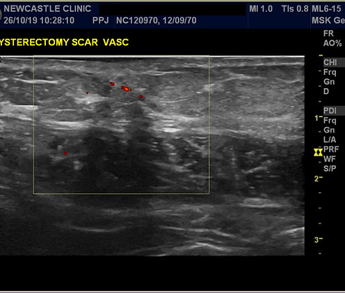
Research 2019
In 2019, we undertook research to evaluate MSTR® using ultrasound imaging.
A General Electric (GE) Soniq S8 ultrasound scanner was used to conduct the test on, in total, twelve test subjects with C-section scars.
Each subject was pre-scanned for:
-
Size and depth of scar tissue
MSTR® work was then applied for a total of 15 minutes per subject, as a single treatment. Immediately after MSTR® treatment each subject underwent a post-treatment ultrasound scan conducted by Consultant Radiologist, Dr Pedada Raju.
These images show pre and post treatment scanned image results.
Note:
-
The size of the scar was measured and displayed (in centimetres or millimetres) in the bottom LEFT of the image.
-
The time and date of the scan is shown toward the top left of the image.
-
For uniformity and to help eliminate unwanted variables, the same practitioner (Alastair McLoughlin) was used to treat all 12 subjects.